NAMNAM ConsultantsUrology OPD · Imphal, Manipur
Home › Kidney stone treatment
Kidney Stone Care · Imphal

Kidney stones,
treated gently.

From medicines that help a small stone pass, to laser surgery through the natural passage with no cut — here is what each option involves, explained in plain language.

● URS & RIRS — no external cut ● Thulium Fibre Laser ● PCNL for large stones ● Stent removal in the OPD
Dr. Nameirakpam Shantajit
Dr. Nameirakpam ShantajitMBBS (SPMC Bikaner) · MS Surgery & M.Ch Urology (RIMS Imphal) · MMC Reg. No. MNP 150
Watch · Manipuri & English RIRS explained by Dr. N. Shantajit

RIRS and PCNL explained by Dr. Shantajit in his own voice, with animation and real images from surgery. Tap to watch ↓

No cutURS & RIRS work through the natural urinary passage
Up to ~2 cmKidney stones usually suited to RIRS
Short stayOften about one day for URS / RIRS
OPDDJ stent removed by flexible cystoscopy, no re-admission
On this page — jump to
Symptoms

How kidney stones show up

Many stones are found on a routine ultrasound without any symptoms. When they do cause trouble, these are the common signs.

⚡

Sudden severe pain

In the side or back, often moving to the lower belly or groin, coming in waves.

🩸

Blood in urine

Pink, red or brown urine — sometimes only seen on a urine test.

🔥

Burning or urgency

Frequent, urgent or painful urination, especially when a stone is near the bladder.

🤢

Nausea & vomiting

Often comes along with the pain of a stone moving down the ureter.

🔁

Repeated infections

Stones can hold bacteria and cause urine infections that keep coming back.

🌡️

Fever with pain

A blocked, infected kidney is an emergency — go to hospital straight away.

Diagnosis

Finding the stone, precisely

The size, position and hardness of the stone decide the right treatment — so the first visit is about getting a clear picture.

Consultation & ultrasound

History, examination and an ultrasound (USG KUB) in the clinic during the same visit. Bring any old scans and reports.

Urine & blood tests

Urine test for infection and blood tests including kidney function (creatinine), done in the in-house laboratory.

CT scan (NCCT KUB)

When a procedure is being considered, a non-contrast CT shows the exact size, position and density of every stone.

A rough guide: stone size and the usual treatment

Every patient is different — stone position, hardness and kidney anatomy also matter.

MedicinesSmall ureteric stones, usually under 5–6 mm
URS / RIRSUreteric stones, and kidney stones up to about 2 cm
PCNLLarge (over ~2 cm), hard or complex kidney stones
0 mm~2 cmlarger

Small stones that pass on their own still need a follow-up scan to confirm they have gone.

Treatment Options

Choosing the right procedure

Your options are explained at the consultation, along with the expected hospital stay and an estimate of cost. Procedures are done at Raj Medicity Hospital, Imphal.

▶ Pre-stented RIRS explained by Dr. Shantajit · in his own voice · Manipuri & English
No cut

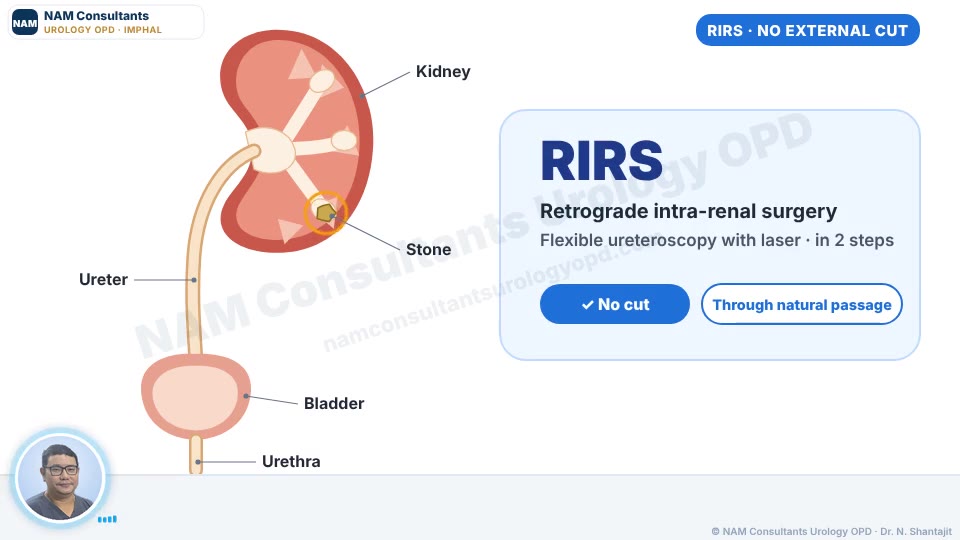
RIRS — Flexible ureteroscopy

Done about 2 weeks after a DJ stent is placed (pre-stenting). A thin flexible scope goes up through the natural urinary passage into the kidney. The laser breaks the stone into fine dust and tiny pieces, which are sucked out through a FANS suction sheath — this keeps the view clear and the pressure inside the kidney low. Usually chosen for kidney stones up to about 2 cm.

Spinal / general anaesthesiaOften ~1 day stayDJ stent usually placed
▶ PCNL explained by Dr. Shantajit · in his own voice · with real surgery images · Manipuri & English
Keyhole

PCNL — Percutaneous nephrolithotomy

Through a small keyhole in the back, a scope (nephroscope) reaches the kidney directly. The stone is broken with a pneumatic lithotripter (or laser) and the pieces are removed through the sheath. Generally preferred for large (over about 2 cm), hard or complex stones, including staghorn stones.

General / spinal anaesthesiaUsually 2–3 days staySmall dressing on the back
No cut

URS — Ureteroscopy

For stones stuck in the ureter (the tube between kidney and bladder). A thin scope reaches the stone through the natural passage; it is broken with laser and removed.

Spinal / general anaesthesiaOften ~1 day stay
No surgery

Medicines & observation

Small stones in the ureter often pass on their own with plenty of fluids, pain relief and medicines that relax the ureter. A repeat scan confirms the stone has passed.

OPD treatmentFollow-up scan
Thulium Fibre Laser machine at the operation theatre, used for kidney stone surgery
Laser Technology

Thulium Fibre Laser

Thulium Fibre Laser (TFL) is the laser platform used at this practice for stone surgery — in RIRS, URS and PCNL.

  • Breaks stones into small fragments or very fine dust, which is sucked out through the suction sheath or passes out with urine
  • Works through a very thin fibre, so it suits flexible scopes inside the kidney
  • Used for stones of different hardness, depending on the case
  • Whether a laser procedure suits you depends on your CT scan — discussed at consultation
Before RIRS

Pre-stenting: why the stent comes first

At NAM Consultants, RIRS is planned in two steps: a DJ stent is first placed about 2 weeks before the laser surgery. It is a short procedure that prepares the ureter, so the main RIRS can be done more safely and smoothly.

Diagram of a DJ (JJ) stent: a soft tube with one curl in the kidney and one in the bladder, running along the ureter past a ureteral stone A DJ (JJ) stent — a soft tube with one curl in the kidney and one in the bladder, holding the ureter open.
↔️

A narrow ureter

Sometimes the ureter is too tight for the scope to pass safely. A stent left for about 2 weeks gently widens it on its own (passive dilatation), so the scope can go up without force.

🦠

Infection with a blocked kidney

If a stone is blocking an infected kidney, the urine must be drained first. The stent relieves the blockage; stone surgery is done once the infection has cleared.

💥

Severe pain or kidney strain

A stent can relieve a blocked kidney quickly — for uncontrolled pain, or when kidney function is affected — while the stone is dealt with in a planned way.

🛡️

Safer, smoother RIRS

A relaxed, wider ureter lowers the risk of injury to the ureter and makes it easier to reach and clear the stone at the main procedure.

What the two-step plan looks like

1
Stent placedShort procedure under anaesthesia, usually same-day
2
About 2 weeksUreter relaxes and widens; infection treated if present
3
RIRSLaser surgery through the prepared ureter
4
Stent removalUsually a few weeks later in the OPD by flexible cystoscopy
Why plan it this wayA pre-stented ureter is already relaxed and wider on the day of RIRS, so the suction sheath and scope pass without force — rather than stretching a tight ureter.
Good to knowIt means two procedures instead of one, and stent symptoms (frequency, mild pain, blood-tinged urine) while it is in. The reasons in your case are explained before you decide.
After Surgery

Recovery & follow-up

🩹

Living with a DJ stent

Mild side or bladder discomfort, frequent urination and blood-tinged urine are common while the stent is in, and settle after removal. Drink plenty of fluids unless told otherwise.

📅

Remove it on time

The stent must come out on the advised date — a forgotten stent can get blocked or encrusted. The WhatsApp chatbot sends removal reminders.

🔭

Removal in the OPD

Done with flexible cystoscopy in a few minutes, without re-admission. Follow-up scans check that the kidney is clear.

Prevention

Keeping stones from coming back

Without prevention, stones come back in about half of patients within 5–10 years. A few daily habits lower that risk.

Stone types

Not all stones are the same

Knowing what your stone is made of is the key to preventing the next one. That is why every removed or passed stone should be sent for stone analysis.

Calcium oxalate

The most common — about 7 in 10 stones

Linked to low fluids, high salt, high-oxalate foods and low citrate.

🩻 Shows on X-ray

Calcium phosphate

About 1 in 10

Linked to alkaline urine, high calcium in urine, or kidney tubule problems (RTA).

🩻 Shows on X-ray

Uric acid

About 1 in 10

Linked to acidic urine, gout, diabetes, obesity and high-purine food. Can often be dissolved with medicine.

🩻 Not seen on plain X-ray — CT needed

Infection (struvite)

Fewer than 1 in 10

Form with certain urine infections and can grow into large "staghorn" stones. Need complete removal.

🩻 Shows on X-ray

Cystine

Rare — 1 to 2 in 100

An inherited condition, often starting young. Needs lifelong prevention.

🩻 Faint on X-ray

💡 Passed a stone at home? Strain your urine through a tea strainer, dry the stone and bring it to the clinic for analysis.

Stone risk check

Are you at high risk of another stone?

Without prevention, about half of stone patients form another stone within 5–10 years. Guidelines identify people at higher risk who need detailed tests. Tick what applies to you.

Medical risk features

Everyday habits

Based on the high-risk stone former criteria in European Association of Urology (EAU) guidelines. For general information only — not a diagnosis. In the clinic, validated scores such as the ROKS nomogram can also estimate the chance of a repeat stone.

💧 How much should you drink?

Very palePale yellow ✓YellowDark yellowAmber — drink more
Aim for pale-yellow urine — at least 2–2.5 litres of urine a day.

People with heart or kidney failure, or on fluid restriction, should follow their own doctor's advice on fluids.

Tests for recurrent stone formers

Finding out why you make stones

Every stone patient needs a few basic tests. People with recurrent stones or other high-risk features need a full metabolic evaluation — it shows exactly what in the urine or blood is causing the stones, so prevention can be specific to you.

Every stone patient

Basic evaluation

  • Stone analysis — what the stone is made of
  • Blood: creatinine (kidney function), calcium, uric acid, sodium, potassium
  • Urine routine: pH, infection, crystals; culture if needed
  • Imaging: ultrasound, and CT KUB when needed
Recurrent / high-risk

Full metabolic evaluation

  • Everything in the basic evaluation
  • Two 24-hour urine collections on your usual diet
  • Parathyroid hormone (PTH) and vitamin D if blood calcium is high or high-normal
  • Blood bicarbonate / gas if renal tubular acidosis is suspected
  • Urine pH profile, and a cystine test if suspected

What a 24-hour urine test measures

MeasuredUsual targetIf abnormal — what helps
Urine volumeMore than 2.5 L a dayLow volume is the commonest risk — drink more
CalciumUnder about 250 mg (women) / 300 mg (men) a dayHigh: less salt, normal dietary calcium; medicines (thiazides) if needed
OxalateUnder about 40 mg a dayHigh: take calcium with meals, limit spinach, nuts, strong tea; check bowel causes
CitrateMore than about 320 mg a dayLow: citrus fruits; potassium citrate medicine
Uric acidUnder about 750–800 mg a dayHigh: less red meat, fish and organ meat; medicine (allopurinol) in some
Sodium (salt)Under about 150 mmol a day — lower is betterHigh salt pulls calcium into the urine
MagnesiumMore than about 3 mmol a dayLow magnesium removes a natural stone blocker
Urine pHAround 6 (target depends on stone type)Very acidic: uric acid stones; high pH: phosphate or infection stones

Approximate targets used in stone clinics; reference values vary between laboratories and are interpreted with your stone type.

How to collect a 24-hour urine sample

1

Start in the morning

Pass your first morning urine into the toilet as usual and note the time — the collection starts now.

2

Collect every drop

For the next 24 hours, collect all urine into the container from the lab. Missing even one sample spoils the result.

3

Finish next morning

At the same time next morning, pass urine into the container — this is the last sample.

4

Keep it cool, bring it in

Keep the container cool (as the lab advises) and bring it to the lab the same morning.

Before you collect
  • Eat and drink as you usually do — so the test shows your real pattern
  • Wait about 3–4 weeks after passing a stone or stone surgery
  • Don't collect during a urine infection or with a stent/catheter, unless told to
  • Usually two collections are done on different days
After the results
  • Prevention is planned for your stone type and test results
  • Tests are repeated a few months after starting treatment to check it is working
  • Regular ultrasound follow-up catches new stones early, while small

Prevention by stone type

Calcium oxalate

Fluids, less salt, normal calcium with meals, citrus; thiazide or potassium citrate when tests show the need.

Calcium phosphate

Fluids, treat high urine calcium; check for parathyroid problems and renal tubular acidosis.

Uric acid

Make urine less acidic (potassium citrate) — this can dissolve many uric acid stones without surgery; allopurinol if uric acid is high.

Infection (struvite)

Remove the stone completely, treat the infection, and keep urine infection-free.

Cystine

Very high fluids (3.5 L or more), alkaline urine, and specific medicines if needed — lifelong follow-up.

🚑

Go to the nearest hospital emergency if you have

  • Fever with chills along with a kidney stone or DJ stent
  • Severe pain not relieved by medicines, or with repeated vomiting
  • Very little or no urine
  • Heavy bleeding in urine or passing clots
WhatsApp numbers and the chatbot are not emergency services.
Questions

Frequently asked questions

Can kidney stones be removed without a cut?

Many can. Small stones often pass with medicines and fluids. URS and RIRS remove stones through the natural urinary passage using a thin scope and laser, with no external cut.

RIRS or PCNL — which one do I need?

It depends mainly on the size, position and hardness of the stone and on your kidney anatomy. RIRS is usually chosen for kidney stones up to about 2 cm; PCNL is generally preferred for larger or complex stones. The choice is made after reviewing your CT scan.

Why do I need a stent before RIRS?

Not everyone does. A stent placed about 2 weeks before RIRS is advised when the ureter is too narrow for the scope to pass safely, when a stone is blocking an infected kidney, or to relieve severe pain or strain on the kidney. It widens the ureter gently and makes the main surgery safer and smoother.

How long is the stent kept before RIRS?

Usually about 2 weeks. If it was placed for an infection, RIRS is done once the infection has been treated and the urine is clear.

Will I need a DJ stent after the surgery too?

Often, yes, after URS, RIRS or PCNL — it lets urine drain while the ureter heals. It is removed in the OPD by flexible cystoscopy on the advised date.

How soon can I go back to work?

Most people return to desk work within a few days after URS or RIRS, and in one to two weeks after PCNL. Heavy physical work may need longer — this depends on the procedure and your recovery.

Do stones come back?

They can — in about half of patients within 5–10 years without prevention. Enough fluids, less salt, normal calcium intake and follow-up scans lower the risk.

What does stone treatment cost?

Costs depend on the procedure, stone size and hospital stay. You will get an estimate after the consultation and tests. For current charges, WhatsApp the clinic at +91 81328 57130.

Am I a high-risk stone former?

You may be if you have had more than one stone, your first stone was in childhood, a close relative has stones, you have one kidney, stones in both kidneys, or conditions such as gout, diabetes, bowel disease or a parathyroid problem. High-risk stone formers need a full metabolic evaluation including 24-hour urine tests.

What is a 24-hour urine test for stones?

You collect all your urine for 24 hours on your usual diet. The lab measures volume, calcium, oxalate, citrate, uric acid, sodium, magnesium and pH. The results show why you are forming stones so prevention can be targeted. Usually two collections are done, about 3–4 weeks after a stone episode or surgery.

Why is stone analysis important?

Different stones need different prevention. Calcium oxalate, uric acid, infection and cystine stones are each prevented in a different way — so knowing the type is the first step.

Can uric acid stones dissolve without surgery?

Often, yes. Making the urine less acidic with medicine (potassium citrate) and drinking plenty can dissolve many uric acid stones, provided the kidney is not blocked or infected.

Should I stop milk and curd to prevent stones?

No. Normal amounts of calcium from milk, curd and paneer, taken with meals, actually lower the risk of calcium oxalate stones. Calcium supplements are different and should be taken only if advised.

Book a consultation

NAM Consultants, Singjamei: mornings daily 6:00–9:30 AM (Mar–Oct) / 6:30–9:30 AM (Nov–Feb), evenings 4–6 PM Mon–Sat · Raj Medicity Hospital: Mon–Sat 11 AM–1 PM

Medically reviewed by Dr. Nameirakpam Shantajit, M.Ch Urology (MMC Reg. No. MNP 150) · Last reviewed: 10 October 2026

Just say "Hi" on WhatsApp+91 70850 51135 · Chatbot 24/7 · Book & queries Clinic WhatsApp81328 57130